Overview
Native space analysis initially was developed for resting state fMRI data. However, it has since expanded to task-based fMRI analysis as well. Native space analyses circumvent the required spatial normalization and smoothing steps in studies of group comparisons. It requires a reconstructed structural MRI image to extract the localization data for each ROI. The inter-modal intra-subject rigid-body registration is done by FSL and Freesurfer epi_reg tools, which are capable of taking into account the EPI field map if it is available.
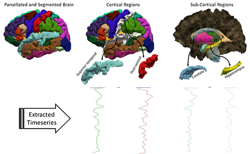
The figure to the right illustrates the technique, in which voxels within each ROI are averaged to generate an ROI-based times series. The current version of the code is works with Freesurfer ROI’s, but is able to also work with any other set of ROI’s.

The figure above shows the flowchart of the overall processes used for native space analysis of the resting state fMRI data. The development of the code for this technique is completed, and available upon request.
Related Publication
Q. R. Razlighi, J. Steffener, C. Habeck, A. Laine, and Y. Stern, “Resting State Inter and Intra Hemispheric Human Brain Functional Connectivity,” Proceedings of IEEE Engineering in Medicine and Biology Society, pp. 6522-6525, Osaka, Japan, Jul. 2013.
Q. R. Razlighi, C. Habeck, J. Steffener, Y. Gazes, L. B. Zahodne, A. M. Brandt, and Y. Stern, “Unilateral disruptions in the default network with aging in native space,” Brain and Behavior , vol. 4, no. 2, pp. 143-157, 2014.
S. Blum, C. Habeck, J. Steffener, Q. R. Razlighi, and Y. Stern, “ Functional connectivity of the posterior hippocampus is more dominant as we age,” Cognitive Neuroscience , vol. 5, no. 3, pp. 150-159, 2014.
Software Released
Please contact Dr. Razlighi for the code and instruction