Overview
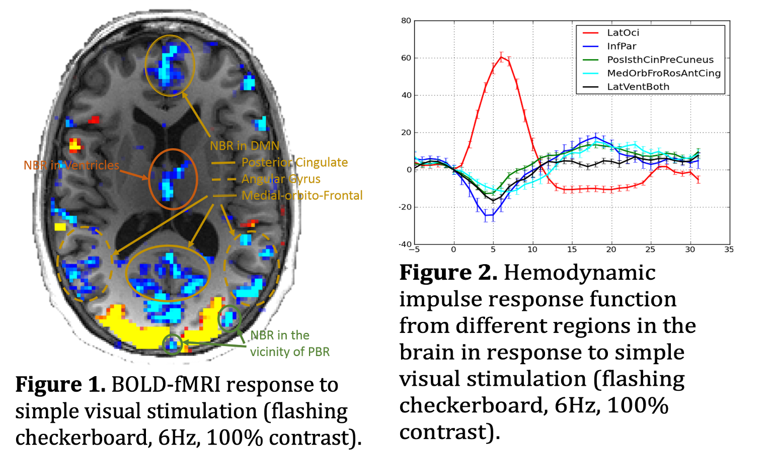
It is puzzling that the human brain’s response to an external stimulus is often more negative than positive when measured using BOLD-fMRI (See Figure 1). Therefore, it is challenging to have a clear understanding of how the brain functions without first delving more deeply into this intriguing phenomenon. The overarching goal of my research is to investigate the mechanisms (either as neuronal, glial, neurophysiological, or neurovascular) that underlie the negative BOLD response (NBR) and the roles and significances of these mechanisms in various brain functions. More importantly, leveraging NBR as an additional functional neuroimaging tool makes it possible to answer some of the key questions in the field about the normal functioning of the human brain, as well as pathological malfunctioning during various neurological and psychiatric diseases.
The main research project in QNL is to investigate the neural and neurophysiological mechanisms underpinning negative BOLD response (NBR). Although there is emerging evidence that sheds light on the mechanism underlying task-based positive BOLD response, the accompanying NBR is mostly unknown (Bentley et al., 2016; Hayden et al., 2009; Mullinger et al., 2014), even though studies investigating NBR started as early as the introduction of the BOLD-fMRI (Shmuel et al., 2002; Smith et al., 2004). One reason for this lack of progress is the possibility that brain regions exhibiting NBR for a specific task may have separate underlying mechanisms. For instance, the NBR often detected in the vicinity of the PBR has mostly been explained by hemodynamics of the neurovascular system rather than by underlying neuronal activity (Hu and Huang, 2015), whereas NBR often detected in the brain DMN regions has been associated with suppression of neuronal activity (Bentley et al., 2016; Hayden et al., 2009). We categorize NBR into four different groups based on its possible underlying mechanism; 1) NBR in the vicinity of the PBR (Hu and Huang, 2015), 2) NBR in the contralateral hemisphere (Smith et al., 2004), 3) NBR in the DMN regions (Hayden et al., 2009), 4) NBR in the ventricles (Bright et al., 2014; Thomas et al., 2013). We feel this categorization is important, as each type of the NBR likely has a distinct underlying mechanism.
Interested Participants
Participant's Instructions
![]() Participant Instructions for FDG Visit (usually the first visit)
Participant Instructions for FDG Visit (usually the first visit)
![]() Participant Instructions for Tau Visit (usually the second visit)
Participant Instructions for Tau Visit (usually the second visit)
![]() Participant Instructions for Amyloid Visit (usually the third visit)
Participant Instructions for Amyloid Visit (usually the third visit)
Related Publication
Parker DB, Razlighi QR. 2019. Task-evoked Negative BOLD Response and Functional Connectivity in the Default Mode Network are Representative of Two Overlapping but Separate Neurophysiological Processes.. Sci Rep. 9(1):14473.
Razlighi QR. 2018. Task-Evoked Negative BOLD Response in the Default Mode Network Does Not Alter Its Functional Connectivity.. Front Comput Neurosci. 12:67.
Funding Sources
This project is completely funded with national institute of aging through an R01 grant.